The OMA is launching a public campaign that recognizes the contributions made by doctors in the health-care system. Starting May 1, the public will be invited to celebrate their doctors by submitting a thank-you note online or by tweeting #thanksdoc.
Share On Social Media
In January 2014, the FDA issued a warning to consumers and physicians about over the counter wart removers being flammable, with multiple reports of fires being started and a variety of injuries to people. We generally discourage the use of these products as we have also had patients with a variety of injuries, and more commonly complaints that the product doesn’t seem to work very well.
Share On Social Media
Keratosis pilaris is a common skin condition that causes rough patches and small, acne-like bumps, usually on the outer arms, and less commonly on the shoulders, thighs, cheeks and buttocks. Keratosis pilaris bumps are either white or red, and don’t hurt or itch. Keratosis pilaris can be frustrating because it’s difficult to treat. However, keratosis pilaris isn’t serious and usually reduces as we age. In the meantime, various creams and self-care measures can improve the appearance of keratosis pilaris.
Keratosis pilaris can occur at any age, although it’s particularly common in children and becomes most noticeable around puberty. Signs and symptoms include:
Small white or red bumps, typically on the upper outer arms, legs, shoulders, buttocks or cheeks
Dry, rough and occasionally itchy skin in the areas with bumps
Worsening in winter, when humidity is low and skin tends to be drier
Keratosis pilaris may be limited to individual, sandpaper-like bumps resembling goose flesh. In some cases, the bumps may become inflamed and cause scarring, especially on the face.
Keratosis pilaris results from the build-up of keratin — a hard protein that protects your skin from harmful substances and infection. The keratin forms a scaly plug that blocks the opening of the hair follicle. Usually many plugs form, causing patches of rough, bumpy skin, often referred to as “chicken skin”.
Why keratin builds up is unknown. It may occur in families for genetic reasons or commonly in the presence of other skin conditions, such atopic dermatitis (genetic eczema). Keratosis pilaris also occurs in otherwise healthy people. Dry skin tends to worsen this condition.
Among the ways to treat keratosis pilaris, the most effective are:
Topical exfoliants:
Medicated creams containing alpha-hydroxy acid, lactic acid, salicylic acid or urea moisturize and soften dry skin while helping to loosen and remove dead skin cells. Depending on their strength, certain creams are available over-the-counter and others require a prescription. Your skin specialist can advise you on the best option for your skin. We like to recommend Alyria Resurfacing Body Cream, with 15% glycolic acid, but also contains hydrating moisturizers to exfoliate and soften the skin at the same time.
Topical retinoids:
Derived from vitamin A, retinoids work by promoting cell turnover and preventing the plugging of the hair follicle. Retinoids may be an effective treatment, but they can cause bothersome skin irritations, such as dryness, redness and peeling. Tretinoin (e.g. Retin-A) and tazarotene (Tazorac) are examples of topical retinoids by prescription. ZO Skin Health has a body cream with 0.05% pure, stable retinol that again, hydrates as it exfoliates and is less irritating than the prescription retinoids.
Laser therapy:
Certain types of keratosis pilaris involving severe redness and inflammation have been successfully treated with laser therapy. Laser treatment involves passing intense bursts of light into targeted areas of skin. This type of treatment may require repeat sessions over the course of a few months, depending on your response. The Profractional laser is able to break up the skin in the affected areas, thus eliminating the plugs; maintenance treatments with laser may be required.
Using a medication regularly may improve the appearance of your skin. But if you stop, the condition returns. And even with medical treatment, keratosis pilaris tends to persist for years. Fortunately people slowly outgrow this annoying skin condition, but it is good to know that there is treatment for its appearance in the meantime.
~ Sheri Roselle, Medical Esthetician at Toronto Dermatology Centre
Share On Social Media
I have had several friends and acquaintances ask me at recent functions whether there is a cost for them or their child to see me for a dermatology appointment. Many of them seem surprised that there is no cost, that seeing a dermatologist in Ontario is an OHIP-covered service, so that anyone requiring expert skin, hair or nails care can see a dermatologist at no cost. That said, for medical concerns about the skin, a referral from a family doctor (GP) or any walk-in clinic or any doctor for that matter, is required to see a dermatologist.
Share On Social Media
It seems that anything and everything can trigger a breakout. The jury is still out on exactly how acne is related to stress, what we order for lunch or simply our genes, but we do know that acne is individual and there is no one-size-fits-all cure. The good news is treating it at any age is getting easier. And while a conversation between you and your dermatologist is your best bet when deciding on treatment, it`s never a bad idea to educate yourself on this common skin condition. Our very own Dr. Anatoli Freiman’s expertise is featured in the Glow magazine as he discusses skin care for acne. Click here to view the article.
Do you remember when you were little our moms and dads would shoo us outside to play all day? They would tell us the fresh air and sunshine was good for us. They would send us off and tell us to be home for dinner or before it got dark. Times have definitely changed! Our parents didn’t know that we needed to be protected from the sun, and of course back then the sun wasn’t nearly as strong as our now receding ozone layer has made it.
The results of that lack of sun protection is now starting to show up on adults in the form of freckles, melasma, fine lines, wrinkles, leathery skin, mottled or discoloured skin, pre-cancers (called “actinic keratoses”) and skin cancers. We are aging before our time. And we all wonder how this could be, because a lot of us have been very good at wearing our sunscreen every morning for the last 10-15 years. Unfortunately the damage has already been done. By all means, protecting our skin now is helping to reduce the damage that could accumulate if we hadn’t been wearing sunscreen for the last 10-15 years, but what can we do about the past?
There is not one perfect solution. However, at Toronto Dermatology Centre we have come up with a cocktail combination of treatment options plus home care that works incredibly well for our patients. Although each patient is individual and their solution may vary, let me tell you about a wonderful treatment plan that is sure to help reverse the signs of photo-damaged skin.
To start, I make sure my patient is using appropriate medical grade home care to get their skin into optimal performance mode. Hydration to the skin is needed for the skin to recover after any service. A serum or moisturizer containing hyaluronic acid will help retain the integrity of the moisture barrier on the skin. Reparative tools such as growth factors, retinol or collagen along with antioxidants increase the healing process and aid in creating new skin that is healthy and rejuvenated.
Once the skin is ready to begin the treatment, my patient will come in to the clinic to have both the BBL (broadband light) and a Profractional laser resurfacing treatment performed on the same day. The BBL will achieve a lifting of the pigmented lesions on the face as well as alleviate broken blood vessels. Immediately after this is done, I will either go straight into a light pass of Profractional over the entire face, getting close up under the eyes as well, or I will apply a numbing cream to the face if the Profractional needs to be treated a little deeper. The Profractional laser eliminates older cells in the dermis of the skin and stimulates new cell creation to replace the old ones.
There are about two days that I consider downtime; however skin may be pink afterwards for up to a week for some patients. Makeup can be worn from day 3 on. The healthier the skin is to start with, the better the results will be. Skin will appear less pigmented, smoother, more glowing and fine lines will be less visible, even after 1 treatment. At this point, it may be advisable for the patient who also has loose or sagging skin to consider a volumizer for the cheek area, such as Juvederm or Restylane, if appropriate.
Although it may take 2-3 BBL/ Profractional treatments for optimal results, it will only take 1 treatment before friends and family members start to see the new you. For those we have treated with this amazing combo therapy so far, the results are undeniable. For minimal downtime, there is no other service that I have seen with such drastic results.
Maybe we can’t change the past, but we can certainly reverse the effects it has had on us! If you grew up at a time when I did, come into Toronto Dermatology Centre and let us help you kick sun damage to the curb!
~ Sheri Roselle, Medical Esthetician at Toronto Dermatology Centre
Share On Social Media
Did you know that April is Rosacea Awareness Month, and the National Rosacea Society encourages physicians and patients to raise awareness of the disease and its impact on affected individuals.
3 new things of note with rosacea:
1. A new Acne and Rosacea Society of Canada is launching!…stay tuned for more information.
2. There is a new oral once-daily medication (Apprilon) for patients who have papules or pustules (acne-like lesions) with their rosacea.
3. In mid-April there will be the first ever topical anti-redness gel on the market called Onreltea. Applied in the morning (you can put other things over top of it such as sunscreen or makeup), it begins working in 30min, with maximal effect at 3-6 hours, and lasts for about 12 hours.
Share On Social Media
Actinic keratosis (or plural actinic keratoses), also called “solar keratosis “and “senile keratosis,” is a pre-malignant condition of thick, scaly, or crusty patches of skin; most commonly they are pink rough spots that feel like sandpaper. It is most common in fair-skinned people and it is associated with those who are frequently and chronically exposed to the sun, as it is usually accompanied by solar damage (e.g. farmers, construction workers, former lifeguards).
They are considered to be pre-cancerous, since some of them progress to squamous cell carcinoma, so treatment is recommended. Untreated lesions have up to 20% risk of progression to squamous cell carcinoma which can be a danger to your health.
Progressive development of these lesions occurs when skin is exposed to the sun constantly and thick, scaly, or crusty areas appear. The scaly or crusty portion is dry and rough. The lesions start out as flat scaly areas and later grow thicker and sometimes become sore.
An actinic keratosis commonly ranges between 2 and 6 millimetres in size, and is usually pink or red with a fine scale or roughness to it. The lesion may appear on any sun-exposed area, such as the face, ears, neck, scalp, chest, backs of hands, forearms, or lips.
Preventive measures recommended for actinic keratosis are similar to those for skin cancer:
Not staying in the sun for long periods of time without protection (e.g., sunscreen, clothing, hats)
Frequently applying quality sunscreens or sunblocks with SPF ratings greater than 30 and that also block both UVA and UVB light
Wearing sun protective clothing such as hats, long-sleeved shirts, long skirts, or trousers
Avoiding sun exposure during mid-day hours is very helpful because ultraviolet light is the most powerful at that time
Actinic keratoses are treated mainly because they are pre-cancerous, but also there is a cosmetic benefit to removing these lesions as many people find them unsightly and sometimes uncomfortable.
There are several treatments for the condition of actinic keratosis, but most commonly used are:
Cryosurgery, e.g. with liquid nitrogen, by “freezing off” the actinic keratosis. This procedure is performed through your dermatologist at Toronto Dermatology Centre. This is particularly useful if you have just a few actinic keratoses.
Creams, e.g. Imiquimod (Zyclara, Aldara) or Ingenol Mebutate (Picato) or 5-FU (Efudex), which are home therapies for treating larger areas of sun damage (areas of “field damage”). For people with many actinic keratoses or frequently recurring actinic keratoses, this therapy should be considered. These are prescribed by your dermatologist and can be a very useful adjunct to liquid nitrogen.
Photodynamic therapy (PDT): this treatment involves applying a topical medicine, Levulan (ALA) or the newer version, Metvix (MLA). The medication is left to incubate on the affected area for an hour or so before an optical energy force is applied to activate the medicine. At Toronto Dermatology Centre we use the BBL (broadband light) that not only helps to clear the actinic keratosis lesions (and “subclinical lesions” or hidden lesions under the skin), but helps to alleviate brown spots (solar damage), decrease broken blood vessels and stimulate collagen production. Although there is minimal discomfort during the actual treatment, it is advised to avoid sunlight or bright indoor lighting for two days following your session. Generally at least two treatments are required, with the addition of maintenance treatments down the road as determined by your dermatologist.
Repeat treatments or maintenance therapy in subsequent years is commonly required since “the damage was already done” many years prior which creates actinic keratoses. Your dermatologist will let you know if and when you need to repeat photodynamic therapy or home “field therapy” with creams.
Actinic keratosis is very common, affecting half of the global population. It is seen most often in fair-skinned individuals, and prevalence may vary with geographical location and age. People who take immunosuppressive drugs, such as organ transplant patients, are 250 times more likely to develop actinic keratoses (AKs) that may lead to skin cancer. If you feel that you may have AKs, it is important to get in to be assessed by your dermatologist as soon as possible. Early diagnosis and treatment is always the best course of action.
~ Sheri Roselle, Medical Esthetician at Toronto Dermatology Centre
Share On Social Media
Actinic keratosis (or plural actinic keratoses), also called “solar keratosis “and “senile keratosis,” is a pre-malignant condition of thick, scaly, or crusty patches of skin; most commonly they are pink rough spots that feel like sandpaper. It is most common in fair-skinned people and it is associated with those who are frequently and chronically exposed to the sun, as it is usually accompanied by solar damage (e.g. farmers, construction workers, former lifeguards).
They are considered to be pre-cancerous, since some of them progress to squamous cell carcinoma, so treatment is recommended. Untreated lesions have up to 20% risk of progression to squamous cell carcinoma which can be a danger to your health.
Progressive development of these lesions occurs when skin is exposed to the sun constantly and thick, scaly, or crusty areas appear. The scaly or crusty portion is dry and rough. The lesions start out as flat scaly areas and later grow thicker and sometimes become sore.
An actinic keratosis commonly ranges between 2 and 6 millimetres in size, and is usually pink or red with a fine scale or roughness to it. The lesion may appear on any sun-exposed area, such as the face, ears, neck, scalp, chest, backs of hands, forearms, or lips.
Preventive measures recommended for actinic keratosis are similar to those for skin cancer:
Not staying in the sun for long periods of time without protection (e.g., sunscreen, clothing, hats)
Frequently applying quality sunscreens or sunblocks with SPF ratings greater than 30 and that also block both UVA and UVB light
Wearing sun protective clothing such as hats, long-sleeved shirts, long skirts, or trousers
Avoiding sun exposure during mid-day hours is very helpful because ultraviolet light is the most powerful at that time
Actinic keratoses are treated mainly because they are pre-cancerous, but also there is a cosmetic benefit to removing these lesions as many people find them unsightly and sometimes uncomfortable.
There are several treatments for the condition of actinic keratosis, but most commonly used are:
Cryosurgery, e.g. with liquid nitrogen, by “freezing off” the actinic keratosis. This procedure is performed through your dermatologist at Toronto Dermatology Centre. This is particularly useful if you have just a few actinic keratoses.
Creams, e.g. Imiquimod (Zyclara, Aldara) or Ingenol Mebutate (Picato) or 5-FU (Efudex), which are home therapies for treating larger areas of sun damage (areas of “field damage”). For people with many actinic keratoses or frequently recurring actinic keratoses, this therapy should be considered. These are prescribed by your dermatologist and can be a very useful adjunct to liquid nitrogen.
Photodynamic therapy (PDT): this treatment involves applying a topical medicine, Levulan (ALA) or the newer version, Metvix (MLA). The medication is left to incubate on the affected area for an hour or so before an optical energy force is applied to activate the medicine. At Toronto Dermatology Centre we use the BBL (broadband light) that not only helps to clear the actinic keratosis lesions (and “subclinical lesions” or hidden lesions under the skin), but helps to alleviate brown spots (solar damage), decrease broken blood vessels and stimulate collagen production. Although there is minimal discomfort during the actual treatment, it is advised to avoid sunlight or bright indoor lighting for two days following your session. Generally at least two treatments are required, with the addition of maintenance treatments down the road as determined by your dermatologist.
Repeat treatments or maintenance therapy in subsequent years is commonly required since “the damage was already done” many years prior which creates actinic keratoses. Your dermatologist will let you know if and when you need to repeat photodynamic therapy or home “field therapy” with creams.
Actinic keratosis is very common, affecting half of the global population. It is seen most often in fair-skinned individuals, and prevalence may vary with geographical location and age. People who take immunosuppressive drugs, such as organ transplant patients, are 250 times more likely to develop actinic keratoses (AKs) that may lead to skin cancer. If you feel that you may have AKs, it is important to get in to be assessed by your dermatologist as soon as possible. Early diagnosis and treatment is always the best course of action.
~ Sheri Roselle, Medical Esthetician at Toronto Dermatology Centre


 It seems that anything and everything can trigger a breakout. The jury is still out on exactly how acne is related to stress, what we order for lunch or simply our genes, but we do know that acne is individual and there is no one-size-fits-all cure. The good news is treating it at any age is getting easier. And while a conversation between you and your dermatologist is your best bet when deciding on treatment, it`s never a bad idea to educate yourself on this common skin condition. Our very own Dr. Anatoli Freiman’s expertise is featured in the Glow magazine as he discusses skin care for acne.
 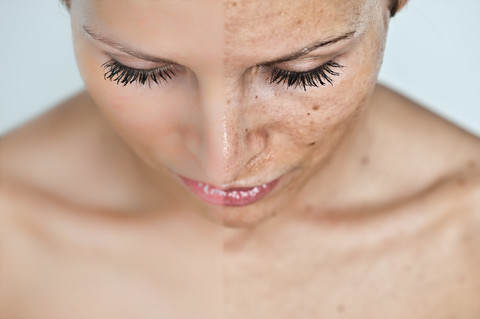 Do you remember when you were little our moms and dads would shoo us outside to play all day? They would tell us the fresh air and sunshine was good for us. They would send us off and tell us to be home for dinner or before it got dark. Times have definitely changed! Our parents didn’t know that we needed to be protected from the sun, and of course back then the sun wasn’t nearly as strong as our now receding ozone layer has made it.
  Did you know that April is Rosacea Awareness Month, and the